
Tarsal Tunnel Syndrome (TTS) / posterior tibial neuralgia
Description of tarsal tunnel syndrome
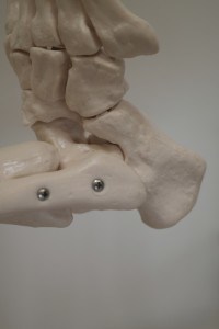
The tarsal tunnel is a channel behind the medial (inner) malleolus (bump on the inside of the ankle) formed by the flexor retinaculum ligament, through which the muscles responsible for bending (flexor muscles) of the toes travel. Also inside the tunnel are blood vessels and nerves supplying the foot with blood and sensibility. In cases where the tendons become irritated, their resulting swilling increases the pressure on the nerve inside the narrow canal.
Symptoms of tarsal tunnel syndrome
Gradually increasing pain under the toes that has burning or stinging characteristics. Most often the pains are located on the inside edge of the foot, but in some cases also in the heel, or in the heel alone.
For half of the patients suffering from TTS the pain radiates along the inside of the lower leg, never reaching knee height. The pain worsens when the patient walks or runs, and subsides at rest. Many experience pain relief from massaging the foot. At night, the foot may feel burning hot and many people also experience restlessness in the calf.
Examination of tarsal tunnel syndrome
- Analysis / inspection / palpation / movement test.
- By pressing or tapping the tibialis posterior nerve behind the innermost malleolus the pain can be produced.
- The pain is exacerbated if a blood pressure cuff on the lower leg is inflated slightly above diastolic pressure for 1 minute.
- Electromyography (EMG) can show increased pressure on the nerve.
Treatment of tarsal tunnel syndrome
- If there is an inward or outward angulation of the heel, this must be corrected this through training, and in the acute phase with shoe inserts.
- Support in running shoes.
- Ultrasound (pulsating), 3 Mhz, 1.5-2 W cm2.
- NSAID for 10 days, possibly as a gel base.
- K-Laser.
- Traumeel ointment or tablets.
In many cases a doctor can administer a single steroid injection into the canal, usually showing positive effect. If the treatments still have had no effect after 3 months, surgical exploration is advised in cleaving the retinaculum. mm. flexorum, and free the nerve and its branches.
Social Medier